Long Bone Diagram Inside : Flashcards - Parts of the Bone - Name #1 & 3 Name #2 ... - There are two types of tissue inside bones:
Long Bone Diagram Inside : Flashcards - Parts of the Bone - Name #1 & 3 Name #2 ... - There are two types of tissue inside bones:. It is found at the ends of long bones, like the head of the femur. Bone anatomy diaphysis epiphysis leg marrow metaphysis trabecular yellow anatomical biology blood body care cartilage cavity compact diagram education educational epiphyseal femoral femur fibula health health. There are two types of tissue inside bones: Cheek bone (zygoma) upper jaw (maxilla). Bone basics and bone anatomyhave you ever seen fossil remains of dinosaur and ancient human bones in textbooks, television, or in person at a museum?
Layer of bone tissue having many small spaces and found just inside the layer of compact bone. Hollow bone or long bone is longer than it is wide and is composed of the following elements the smallest units of bones are found inside the compact bone. Bone basics and bone anatomyhave you ever seen fossil remains of dinosaur and ancient human bones in textbooks, television, or in person at a museum? Parts of a long bone. In adults, red marrow is found primarily in the breastbone, hips, ribs, skull, spinal bones and at the end of long bones of the arms and legs.
Diagram Human Bone Anatomy Useful Education Stock Vector ... from image.shutterstock.com The shiny, articulating cartilage on the ends of a bone. Bone marrow is the soft, highly vascular and flexible connective tissue within bone cavities. There are two main types of marrow, red and yellow. Sectional diagram of a long bone. Long bone growth is similar to endochondral ossification (there's cartilage there just like in development). Rethinking pain education online course: This tissue is made up of smaller plates filled with red bone marrow. Bone basics and bone anatomyhave you ever seen fossil remains of dinosaur and ancient human bones in textbooks, television, or in person at a museum?
The shaft tends to be cylindrical in form.
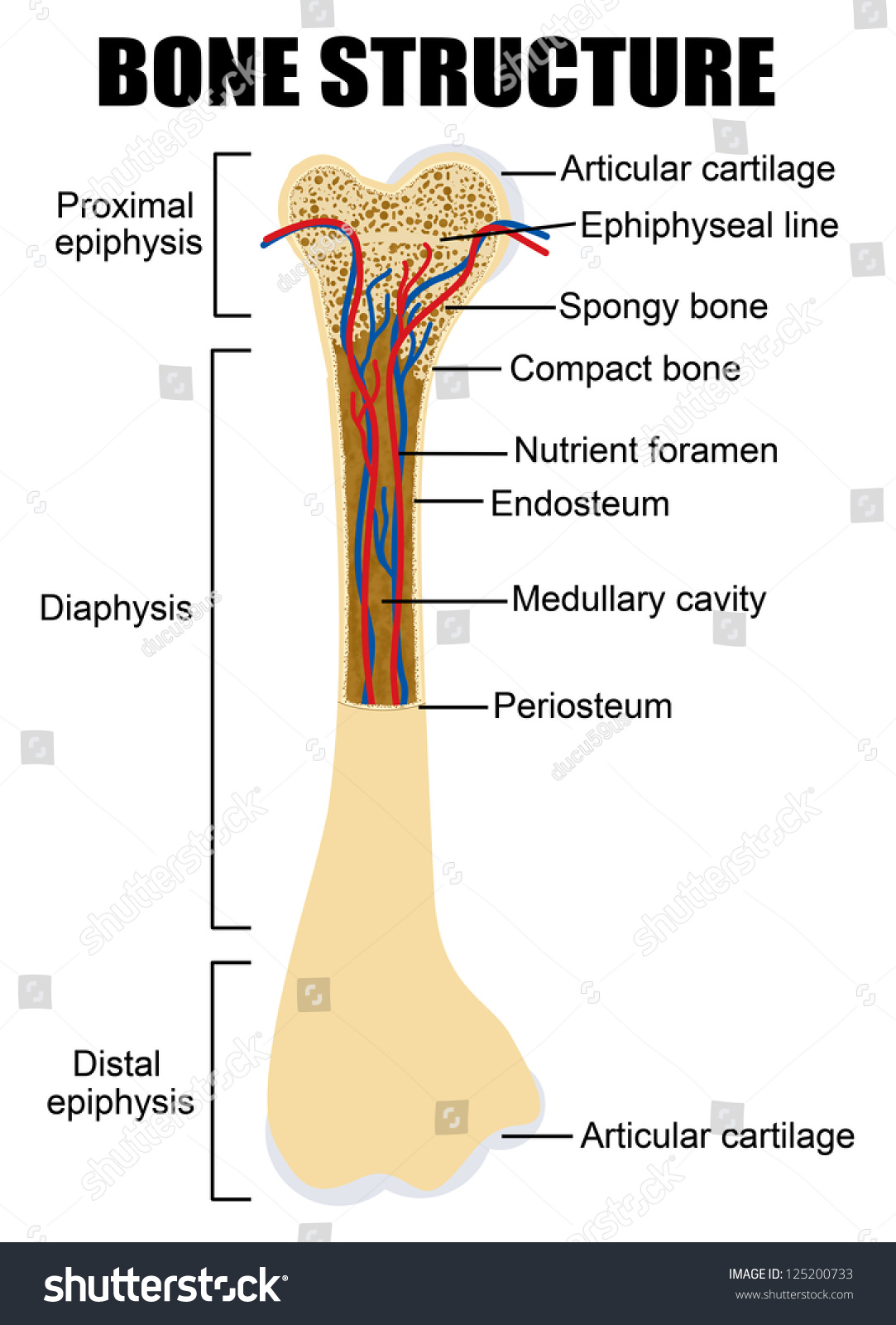
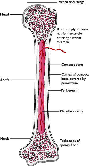
This is called the diaphysis. Inside bone diagram data wiring diagram today. Inside the diaphysis is a tubelike area called the medullary cavity, which houses red marrow during childhood, which is replaced by yellow marrow as a person ages. Bones are mostly made of the protein collagen, which forms a soft framework. Layer of bone tissue having many small spaces and found just inside the layer of compact bone. Long bones grow more than the other classes of bone throughout childhood and so are responsible for the bulk of our height as adults. Bones come in all shapes and sizes and have many roles. Midsection (shaft) of a long bone. The smallest bone in the human body is called the stirrup bone, located deep inside the ear. The outer part of a long bone is made of compact bone. Bone marrow is the soft, highly vascular and flexible connective tissue within bone cavities. Cartilage cells in epiphyseal plate divide, youngest toward epiphysis. The blood vessels inside a bone.
As shown in figure 2. Hollow bone or long bone is longer than it is wide and is composed of the following elements the smallest units of bones are found inside the compact bone. In this article, we explain their function, what they are made of, and the types of cells involved. Bone is found in the shafts of long bone and consists of various cylindrical units named as haversian system 47. The long bones are those that are longer than they are wide.
Chapter 5: Bones - Kinesiology 170 with Maves at ... from classconnection.s3.amazonaws.com As shown in figure 2. The terms osteogenesis and ossification are often used synonymously to indicate the process of bone formation. Long bones, especially the femur and tibia, are subjected to most of the load during daily activities and they are crucial for skeletal mobility. Inside bone diagram data wiring diagram today. Vector illustration for medical, educational and science use. Related posts of long bone diagram labeled human bone parts. Sectional diagram of a long bone. Bone marrow is the soft, highly vascular and flexible connective tissue within bone cavities.
The tough membrane covering the shaft of the bone.
Related posts of long bone diagram labeled human bone parts. 4 looking at the inside of the bone. The long bones are those that are longer than they are wide. Sectional diagram of a long bone. The blood vessels inside a bone. Related online courses on physioplus. Diagram of blood and nerve supply to bone. The mineral calcium phosphate hardens this framework, giving it. Layer of bone tissue having many small spaces and found just inside the layer of compact bone. Bone anatomy diaphysis epiphysis leg marrow metaphysis trabecular yellow anatomical biology blood body care cartilage cavity compact diagram education educational epiphyseal femoral femur fibula health health. Rethinking pain education learn how to teach your patient about their pain powered by physiopedia. Bone basics and bone anatomyhave you ever seen fossil remains of dinosaur and ancient human bones in textbooks, television, or in person at a museum? See more ideas about anatomy, anatomy bones, human anatomy.
Sectional diagram of a long bone. This is called the diaphysis. Inside the diaphysis is a tubelike area called the medullary cavity, which houses red marrow during childhood, which is replaced by yellow marrow as a person ages. There is a printable worksheet available for download here so you can take the quiz with pen and paper. Bone anatomy diaphysis epiphysis leg marrow metaphysis trabecular yellow anatomical biology blood body care cartilage cavity compact diagram education educational epiphyseal femoral femur fibula health health.
Skeletal system | Veterian Key from veteriankey.com In adults, red marrow is found primarily in the breastbone, hips, ribs, skull, spinal bones and at the end of long bones of the arms and legs. The smallest bone in the human body is called the stirrup bone, located deep inside the ear. As shown in figure 2. There is a printable worksheet available for download here so you can take the quiz with pen and paper. Bone anatomy diaphysis epiphysis leg marrow metaphysis trabecular yellow anatomical biology blood body care cartilage cavity compact diagram education educational epiphyseal femoral femur fibula health health. Bones are mostly made of the protein collagen, which forms a soft framework. Through the concentrated arrangement of bone lamellae, several thin long cylinders are formed. Layer of bone tissue having many small spaces and found just inside the layer of compact bone.
Related posts of long bone diagram labeled human bone parts.
It is found at the ends of long bones, like the head of the femur. This is an online quiz called long bone diagram. Bone basics and bone anatomyhave you ever seen fossil remains of dinosaur and ancient human bones in textbooks, television, or in person at a museum? Larger bones contain bone marrow, a spongy tissue inside the bones. Human skeleton, the internal skeleton that serves as a framework for the body. The outer part of a long bone is made of compact bone. In adults, red marrow is found primarily in the breastbone, hips, ribs, skull, spinal bones and at the end of long bones of the arms and legs. Vector illustration for medical, educational and science use. Layer of bone tissue having many small spaces and found just inside the layer of compact bone. There are two main types of marrow, red and yellow. A typical long bone shows the gross anatomical characteristics of bone. See more ideas about anatomy, anatomy bones, human anatomy. It is composed of hematopoietic tissue that has become inactive.
Cartilage cells in epiphyseal plate divide, youngest toward epiphysis long bone diagram. Long bones follow the process of endochondral ossification where the diaphysis grows inside of cartilage from a primary ossification center until it forms most of the bone.